Background & Objectives
Although alpha-synuclein-related pathology is the hallmark of dementia with Lewy bodies (DLB), cerebrovascular and AD pathologies are common in DLB patients. Little is known about the contribution of these pathologies to neurodegeneration in DLB. We investigated associations of cerebrovascular, β-amyloid, and tau biomarkers with gray matter (GM) volume in probable DLB patients.
Methods
We assessed probable DLB patients and cognitively unimpaired (CU) controls with 11C-Pittsburgh Compound-B (PiB) and 18F-Flortaucipir positron emission tomography (PET), as markers of β-amyloid and tau, respectively. MRI was used to assess white matter hyperintensity volume (WMH, a marker of cerebrovascular lesion load) and regional GM volume (a marker of neurodegeneration). We used correlations and ANCOVA in the entire cohort and structural equation models in the DLB patients to investigate associations of WMH volume and regional β-amyloid and tau PET standardized uptake value ratios (SUVr), with regional GM volume.
Results
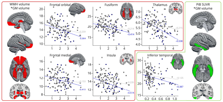
We included 30 DLB patients (69.3±10.2 years old, 87% men) and 100 CU controls balanced on age and sex. Compared to CU, DLB patients showed lower GM volume across all cortical and subcortical regions except for cuneus, putamen, and pallidum. A larger WMH volume was associated with lower volume in the medial and orbital frontal cortices, insula, fusiform cortex, and thalamus in DLB patients. A higher PiB SUVr was associated with lower volume in the inferior temporal cortex, while Flortaucipir SUVr did not correlate with gray matter volume. Structural equation models showed that a higher age and absence of the APOE ε4 allele were significant predictors of higher WMH volume, and WMH volume in turn was a significant predictor of GM volume in medial and orbital frontal cortices, insula, and inferior temporal cortex. In contrast, we observed two distinct paths for the fusiform cortex, with age having an effect through PiB and Flortaucipir SUVr on the one path, and through WMH volume on the other path.
Discussion
Probable DLB patients have widespread cortical atrophy, most of which likely is influenced by alpha-synuclein-related pathology. Although cerebrovascular, β-amyloid, and tau pathologies often coexist in probable DLB, their contributions to neurodegeneration seem to be region-specific.
View Full Article: https://n.neurology.org/content/early/2022/11/28/WNL.0000000000201579