Abstract
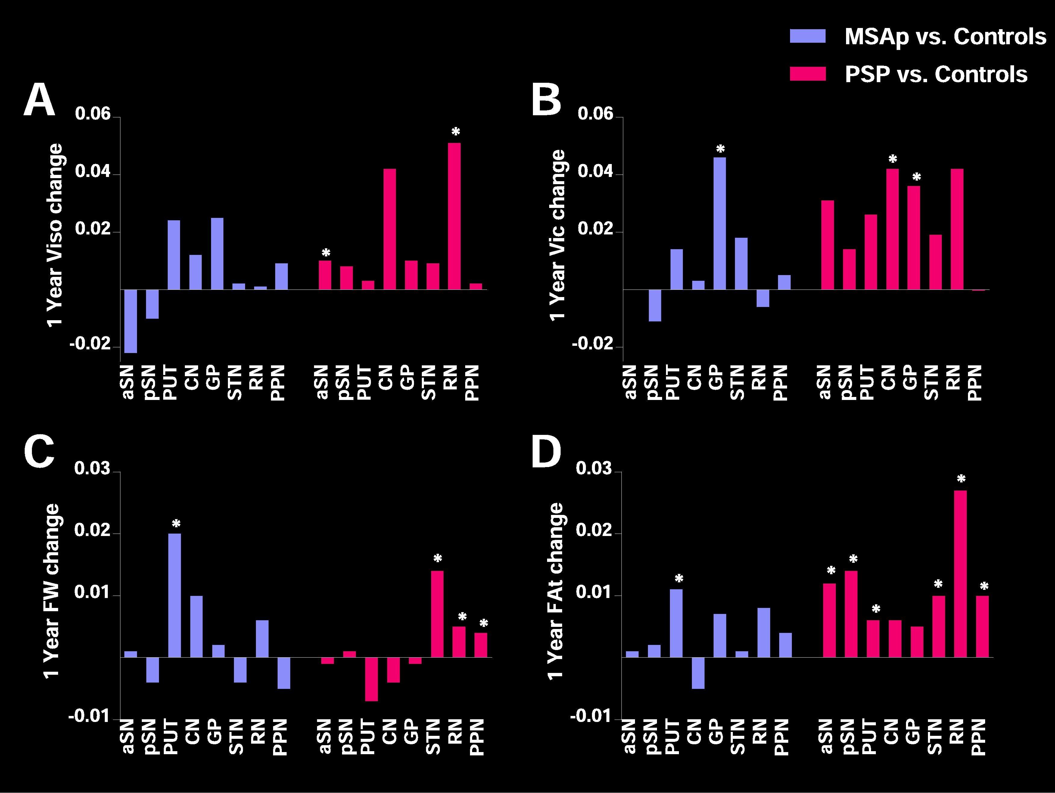
Advanced diffusion imaging which accounts for complex tissue properties, such as crossing fibers and extracellular fluid, may detect longitudinal changes in widespread pathology in atypical Parkinsonian syndromes. We implemented fixel-based analysis, Neurite Orientation and Density Imaging (NODDI), and free-water imaging in Parkinson’s disease (PD), multiple system atrophy (MSAp), progressive supranuclear palsy (PSP), and controls longitudinally over one year. Further, we used these three advanced diffusion imaging techniques to investigate longitudinal progression-related effects in key white matter tracts and gray matter regions in PD and two common atypical Parkinsonian disorders. Fixel-based analysis and free-water imaging revealed longitudinal declines in a greater number of descending sensorimotor tracts in MSAp and PSP compared to PD. In contrast, only the primary motor descending sensorimotor tract had progressive decline over one year, measured by fiber density (FD), in PD compared to that in controls. PSP was characterized by longitudinal impairment in multiple transcallosal tracts (primary motor, dorsal and ventral premotor, pre-supplementary motor, and supplementary motor area) as measured by FD, whereas there were no transcallosal tracts with longitudinal FD impairment in MSAp and PD. In addition, free-water (FW) and FW-corrected fractional anisotropy (FAt) in gray matter regions showed longitudinal changes over one year in regions that have previously shown cross-sectional impairment in MSAp (putamen) and PSP (substantia nigra, putamen, subthalamic nucleus, red nucleus, and pedunculopontine nucleus). NODDI did not detect any longitudinal white matter tract progression effects and there were few effects in gray matter regions across Parkinsonian disorders. All three imaging methods were associated with change in clinical disease severity across all three Parkinsonian syndromes. These results identify novel extra-nigral and extra-striatal longitudinal progression effects in atypical Parkinsonian disorders through the application of multiple diffusion methods that are related to clinical disease progression. Moreover, the findings suggest that fixel-based analysis and free-water imaging are both particularly sensitive to these longitudinal changes in atypical Parkinsonian disorders.
Conclusions
In summary, fixel-based analysis based on a multi-shell scan and free-water imaging based on a single-shell scan were particularly sensitive to widespread longitudinal changes in atypical Parkinsonism. The present results identify novel extra-nigral and extra-striatal longitudinal progression effects in atypical Parkinsonism when applying multiple diffusion methods that are associated with clinical disease progression. Fixel-based analysis and free-water imaging are particularly sensitive to these longitudinal changes in atypical Parkinsonism. These findings are critical for future applications of neuroimaging biomarkers for tracking progression in Parkinsonian disorders, especially atypical Parkinsonian disorders that rely on rapid and early identification for early intervention and clinical trial participation.
Source: https://www.sciencedirect.com/science/article/pii/S2213158222000870?via%3Dihub#s0095